


From the article you will learn the peculiarities of small pelvic varicose veins in women - it is the deformation of the veins in the pelvic region with impaired blood flow to the internal and external genitalia.
General Information
In the literature, small pelvic varicose veins are also referred to as "pelvic constriction syndrome", "varicocele in women", "chronic pelvic pain syndrome". In women. Most often, pelvic venous pathology is diagnosed in patients aged 25-45 years in the reproductive period.
In the vast majority of cases (80%), varicose transformation affects the ovarian veins and is very rare (1%) in the wide ligament veins of the uterus. According to modern medical approaches, treatment of VVMT should be carried out not so much in terms of gynecology, but primarily in terms of phlebology.
Causing pathology
In women with varicose veins of the pelvic organs, doctors understand the change in the structure of the blood vessel walls characteristic of other types of disease - weakening, which is followed by stretching and the formation of "pockets" in which the blood stops. It is extremely rare for only the vessels of the pelvic organs to be damaged. In about 80% of patients with this form is observed varicose veins of the uterine veins, vessels of the lower extremities.
Varicose veins of the small pelvis are most pronounced in women. This is due to the anatomical and physiological characteristics, which indicate a tendency to weaken the venous walls:
- Hormonal fluctuations, including the menstrual cycle and pregnancy;
- Increased pressure in the small pelvis, which is characteristic of pregnancy;
- Periods of more active filling of the veins with blood, including cyclical menstruation, during pregnancy, as well as during sex.
All these phenomena belong to the category of factors provoking varicose veins. And they are found only in women. Most patients experience small pelvic vein varicose veins during pregnancy as a simultaneous layer of provoking factors occurs. According to statistics, varicose veins of the small pelvis in men are 7 times less than among the fairer sex. They have a more diverse set of provoking factors:
- Hypodynamics - long-term maintenance of low physical activity;
- Increased physical activity, especially weight gain;
- Obesity;
- Lack of sufficient fiber in the diet;
- Inflammatory processes in the organs of the genitourinary system;
- Sexual dysfunction or a clear refusal to have sex.
Genetic predisposition can also lead to pathology of the plexuses located inside the small pelvis. According to statistics, varicose veins of the perineum and pelvic organs are most often diagnosed in women whose relatives had this disease. The first changes in them can be observed in adolescence during puberty.
The greatest risk of developing varicose veins of the uterus in women with involvement of the pelvic blood vessels is observed in patients who have venous pathology in other parts of the body. In this case we are talking about congenital weakness of the veins.
Etiopathogenesis
Proctologists believe that the following major causes always contribute to the onset of VVP: valvular insufficiency, venous obstruction, and hormonal changes.
Pelvic venous occlusion syndrome may develop due to the congenital absence or insufficiency of venous valves, which has been revealed by anatomical studies in the last century and this is confirmed by modern data.
It was also found that in 50% of patients varicose veins are genetic in nature. FOXC2 was one of the first identified genes to play an important role in the development of VVP. A link is currently established between disease development and gene mutations (TIE2, NOTCH3), thrombomodulin levels, and type 2 transformer growth factor β. These factors contribute to a change in the structure of the valve itself or the venous wall - all of which lead to a failure of the valve structure; Enlargement of the vein, causing a change in valve function; Progressive to reflux and eventually to varicose veins.
An important role in the development of the disease can be played by connective tissue dysplasia, the morphological basis of which is a decrease in the content of different types of collagen or a violation of the ratio between them, which leads to a decrease in the strength of the veins. .
The frequency of VVP is directly proportional to the amount of hormonal changes that are particularly pronounced during pregnancy. The volume of pelvic veins in pregnant women increases by 60% due to mechanical compression of the pelvic vessels by the pregnant uterus and the vasodilating effect of progesterone. This venous enlargement lasts for up to a month after delivery and can lead to venous valve insufficiency. In addition, during pregnancy, the mass of the uterus increases, its positional changes occur, leading to stretching of the ovarian veins, followed by venous edema.
Risk factors also include endometriosis and other inflammatory diseases of the female reproductive system, estrogen therapy, unwanted working conditions for pregnant women, which include heavy physical labor and prolonged forced position (sitting or standing) during the working day.
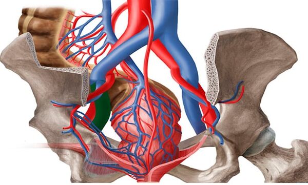
The anatomical features of the outflow from the small pelvic veins also contribute to the formation of varicose veins in the small pelvis. The diameter of the ovarian veins is usually 3-4 mm. The long and thin ovarian veins on the left flow into the left renal vein, and on the right into the inferior vena cava. Normally, the left renal vein is located in front of the aorta and behind the superior mesenteric artery. The physiological angle between the aorta and the superior mesenteric artery is approximately 90 °.
This normal anatomical position prevents compression of the left renal vein. On average, the angle between the aorta and the upper mesenteric artery is 51 ± 25 ° in adults, 45, 8 ± 18, 2 ° in children and 45, 3 ± 21, 6 ° in girls. When the angle is reduced to 39, 3 ± 4, from 3 ° to 14. 5 °, aorto-mesenteric compression, or Macnatuna syndrome, occurs. This is the so-called anterior, or true, Nutcracker syndrome, which has great clinical significance. Posterior Nutcracker Syndrome is rarely seen in patients who have a retroaortic or ring arrangement of the distal left renal vein. Obstruction of the proximal venous bed causes an increase in pressure in the renal vein, leading to renovar reflux in the left ovarian vein with the development of chronic pelvic venous insufficiency.
May-Turner syndrome - Compression of the left common femoral vein by the right common femoral artery - is also one of the etiological factors of pelvic varicose veins. It occurs in no more than 3% of cases, is more common in women. Currently, due to the introduction of radiation and endovascular imaging methods in practice, the detection of this pathology is becoming more frequent.
Classification
Varicose veins are divided into the following forms:
- Primary type of varicose veins: Increased pelvic blood vessels. The cause is 2 types of valvular insufficiency: acquired or congenital.
- The secondary form of pelvic vein thickening is diagnosed exclusively in the presence of gynecological pathologies (endometriosis, neoplasms, polycystic ovaries).
Gradually develops varicose veins of the pelvis. There are several major stages in the development of the disease in medical practice. They differ in the presence of complications and the prevalence of the disease:
- ᲞNir quality. Changes in the structure of the ovarian venous valves can be due to hereditary causes or acquired. The disease is characterized by an increase in the diameter of the veins up to 5 mm. The left ovary has a pronounced enlargement in the outer parts.
- Second degree. This degree is characterized by the spread of pathology and damage to the left ovary. The veins of the uterus and right ovary may also dilate. The diameter of the expansion reaches 10 mm.
- Third degree. The diameter of the veins increases to 1 cm. Enlargement of the veins is observed in the right and left ovaries equally. This stage is due to pathological events of gynecological nature.
It is also possible to classify the disease according to the main reason for its development. There is a primary degree in which the enlargement is caused by defective functioning of the venous valves and a secondary degree which is the result of chronic female diseases, inflammatory processes or complications of an oncological nature. The degree of the disease may vary according to anatomical features, indicating the location of the vascular disorder:
- Intracastal abundance.
- Vulvar and perineal.
- Combined forms.
Symptoms and clinical manifestations
In women, pelvic varicose veins are accompanied by severe but nonspecific symptoms. Often, the manifestations of this disease are considered as signs of gynecological disorders. The main clinical symptoms of pelvic varicose veins in women with pelvic vascular damage are:
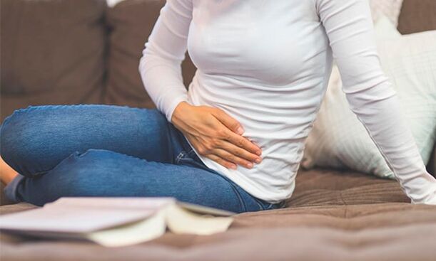
- Non-menstrual pain in the lower abdomen. Their intensity depends on the stage of the venous lesion and the scale of the process. Grade I varicose veins of the small pelvis are characterized by periodic, mild pain that extends to the lower back. In later stages it is felt in the abdomen, perineum and lower back and is long and intense.
- Abundant mucous discharge. The so-called leukorrhea does not have an unpleasant odor, does not change color, which indicates an infection. The volume of the discharge increases in the second phase of the cycle.
- Exacerbation of symptoms of premenstrual syndrome and dysmenorrhea. Even before menstruation, the pain in women increases, leading to difficulty walking. During menstrual bleeding it can become unbearable, spreading to the entire pelvic area, perineum, lower back and even the thighs.
- Another characteristic sign of uterine varicose veins in women is discomfort during sexual intercourse. It is felt in the vulva and vagina and is characterized as a dull ache. This can be noticed at the end of sexual intercourse. In addition, the disease is accompanied by increased anxiety, irritability, and mood swings.
- As in the case of varicose veins in men, the interest in sex in women with such a diagnosis gradually disappears. The cause of the dysfunction is both permanent discomfort and a decrease in the production of sex hormones. In some cases, infertility can occur.
Instrumental diagnostics
Diagnosis and treatment of varicose veins is performed by a phlebologist, a vascular surgeon. Currently, due to new technologies, the number of VVP detections has increased. Patients with CPP are examined in several stages.
- The first stage is a routine examination by a gynecologist: taking a medical history, manual examination, ultrasound examination of the pelvic organs (to rule out other pathologies). According to the results, the examination is additionally prescribed by a proctologist, urologist, neurologist and other related specialists.
- If the diagnosis is unclear but there is a suspicion of VVPT, an ultrasound of the pelvic veins is performed with ultrasound angiography (USAS). It is a non-invasive, highly informative method of screening diagnosis used in all women with suspected VVPT. If previously it was thought that only examination of the pelvic organs was sufficient (venous examination was considered difficult to access and optional), then at this stage ultrasound examination of the pelvic veins is a mandatory procedure. With the help of this method it is possible to determine the varicose veins of the small pelvis by measuring the diameter, the speed of blood flow in the veins and to find out in advance what is the leading pathogenetic mechanism - failure. Ovarian veins or venous obstruction. Also, this method is used for dynamic evaluation of conservative and surgical treatment of VVPT.
- The study is performed transvaginally and transabdominally. Parametric veins, uterine-like plexuses, and uterine veins are visualized transvaginally. VariousAccording to the author, the diameter of the named localization vessel varies from 2, 0 to 5, 0 mm (average 3, 9 ± 0, 5 mm), e. ი. Not more than 5 mm, and the average diameter of arcuate veins is 1, 1 ± 0, 4 mm. Veins larger than 5 mm in diameter are considered dilated. The inferior vena cava, jugular veins, left renal vein, and ovarian veins are examined transabdominally to rule out thrombotic masses and extravascular compression. The length of the left renal vein is 6 to 10 mm and its average width is 4 to 5 mm. Normally, the left renal vein at the site where it passes through the aorta is somewhat flattened, but its transverse diameter is reduced 2-2, 5 times without significant acceleration of blood flow, ensuring normal drainage without increasing the pressure in the aorta. Zone. In the case of venous stenosis against the background of abnormal compression, there is a significant reduction in its diameter - 3, 5-4 times and an acceleration of blood flow - more than 100 cm / sec. The sensitivity and specificity of this method are 78 and 100%, respectively.
- Mandatory examination of the pelvic veins includes examination of the ovarian veins. They are located along the anterior wall of the abdomen, along the abdominal muscle of the rectum, slightly to the lateral femoral veins and arteries. In the US, the sign of ovarian venous insufficiency is considered to be the presence of retrograde blood flow greater than 5 mm in diameter. Ultrasound examination of the lower extremities, perineum, vulva, inner thigh, and gluteal veins should be performed for a complete examination, relapse prevention, and correct treatment tactics.
- The development of medical technology has led to the use of new diagnostic methods. In the third stage, after ultrasound examination of the diagnosis, radiation diagnostic methods are used to confirm it.
- Pelvic phlebography with selective bilateral radiolucent ovarography is one of the radiation invasive diagnostic methods that is performed only in a hospital setting. This method has long been considered the diagnostic "gold standard" for assessing dilatation and diagnosing valvular insufficiency in the pelvic veins. To the ovarian veins. Thus, it is possible to identify anatomical variants of the structure of the ovarian veins, to determine the diameter of the genital and pelvic veins.
- The retrograde contrast of the genital veins at the height of the Valsalva test serves as a pathognomonic angiographic sign of their valvular insufficiency, with visualization of sharp enlargement and tortuosity, respectively. This is the most accurate method for detecting May-Turner syndrome, thrombophlebitic changes in the iliac and inferior vena cava.
- When the left renal vein is compressed, perianal venous collaterals are defined by retrograde blood flow to the gonadal veins, with contrast stagnation in the renal vein. The method measures the pressure gradient between the left kidney and the inferior vena cava. Normally, this is 1 mm Hg. Art . ; The gradient is equal to 2 mm Hg. Art. , May indicate light compression; With gradient >3 mm Hg. Art. Can be diagnosed with aorto-mesenteric compression syndrome with hypertension in the left renal vein and a gradient >5 mm Hg. Art. Is considered to be a hemodynamically significant stenosis of the left renal vein. Determining the pressure gradient is an important element of the diagnosis, since due to its importance, substantially different surgical interventions are planned for the small pelvic veins, which is very important in modern conditions. Currently this study (with a normal pressure gradient) can be used for therapeutic purposes - for embolization of ovarian veins.
- The next radiation method is pelvic vein emission computed tomography in vitro with labeled erythrocytes. Characterized by deposition of labeled erythrocytes in the pelvic veins and visualization of the genital veins. Pelvic veins in the saphenous veins of the legs and perineum. Normally, the ovarian veins are not contrasting, no accumulation of radiopharmaceuticals is observed in the venous plexuses. The coefficient of pelvic venous occlusion is calculated for the objective assessment of the quality of small pelvic venous occlusion. But this method also has disadvantages: invasiveness, relatively low spatial resolution, inability to accurately determine the diameter of the veins, therefore, at present it is not so often used in clinics.
- Video laparoscopic examination is a valuable tool for assessing the undiagnosed. Among other methods, it can help determine the causes of pain and prescribe the right treatment. When varicose veins of the small pelvis dilate in the ovary, along the round and wide ligaments of the uterus, the veins may visually appear as cyanotic, dilated blood vessels, with a thin and tense wall. The use of this method is significantly limited by the following factors: the presence of retroperitoneal adipose tissue, the ability to assess varicose veins only in a limited area, and the inability to determine reflux through the veins. Currently the use of this method is diagnostically justified in case of suspicious multifocal pain. Laparoscopy visualizes the underlying causes of CPP, such as foci of endometriosis or adhesions, in 66% of cases.
Characteristics of therapy
For the full treatment of small pelvic varicose veins, a woman should follow all the recommendations of the doctor, as well as change her lifestyle. First of all, you should pay attention to the loads, if they are too high, they should be reduced, if the patient leads an overly sedentary lifestyle, it is necessary to do sports, walk more often, etc. Sh.
Patients with varicose veins are strongly advised to change their diet, consume as few junk foods as possible (fried, smoked, large amounts of sweet, salty, etc. ), alcohol, caffeine. It is better to give preference to vegetables and fruits, dairy products, cereals.
Also, as a prevention of disease progression and treatment goals, doctors prescribe compression underwear to patients with varicose veins.
Drugs
ERCT therapy involves several important points:
- Escape from reverse venous blood flow;
- Alleviation of disease symptoms;
- Stabilization of vascular tone;
- Improved blood circulation in tissues.
Preparations for varicose veins should be taken in courses. The rest of the drugs, which act as painkillers, are allowed to drink only during a painful attack. For effective therapy, your doctor will often prescribe the following medications:
- Phleboprotectors;
- Enzymatic preparations;
- Medications that relieve inflammatory processes in varicose veins;
- Pills to improve blood circulation.
Surgical treatment
It is noteworthy that conservative treatment methods give truly visible results mainly in the early stages of varicose veins. At the same time, the problem can be solved fundamentally and the disease can be completely eliminated only by surgery. There are many variations of surgical treatment of varicose veins in modern medicine, consider the most common and effective types of surgeries:
- Embolization of veins in the ovaries;
- Sclerotherapy;
- Plastic ligation of the uterus;
- Removal of enlarged veins by laparoscopy;
- Stretching of small pelvic veins with special medical fasteners (clipping);
- Crocectomy - ligation of the veins (prescribed if, in addition to the pelvic organs, the vessels of the lower extremities are damaged).
Only symptomatic therapy of small pelvic varicose veins is possible during pregnancy. If you prefer to wear compression stockings, take phlebotonics on the recommendation of a vascular surgeon. Phlebosclerosis of varicose veins of the perineum may occur in the II-III trimester. If there is a high risk of bleeding during spontaneous delivery due to varicose veins, a choice is made in favor of surgical delivery.
Physiotherapy
The system of physical activity for the treatment of varicose veins in women consists of exercises:
- "Bicycle". We lie on our backs, arms folded or placed across the body. Raising our legs, we make circular movements with them, as if we were riding a bicycle.
- "Vodka". We look face to face on any solid, comfortable surface. Lift your legs up and gently start them behind your head. Hold your waist with your hands and place your elbows on the floor, slowly straighten your legs, lift your body up.
- "რScratel". The starting position is on the back. Raise your closed legs slightly above floor level. Spread the lower limbs on the sides, turn back and repeat.
Possible complications
Why are small pelvic varicose veins dangerous? The following consequences of the disease are often observed:
- Inflammation of the uterus, its appendages;
- Uterine bleeding;
- Disorders of the bladder;
- Development of venous thrombosis (small percentage).
Prevention
In order to make the small pelvic varicose veins disappear as soon as possible and prevent the recurrence of pelvic organ pathology in the future, it is worth following the simple preventive rules:
- Perform gymnastic exercises daily;
- Avoid constipation;
- Follow a diet in which plant fiber should be present;
- Do not stay in one position for long;
- Take a perineum contrast shower;
- To prevent varicose veins from appearing, it is best to wear particularly comfortable shoes and clothing.
Preventive measures aimed at reducing the risk of emergence and progression of small pelvic varicose veins are mainly reduced to a normalization of lifestyle.